
New Technology Redefines UTHealth Anatomy Lab

We all know how to swipe left, right, up or down. Pinching the screen to zoom in or out just seems basic at this point. And our opposable thumbs are physical adaptations that have given us the evolutionary advantage to text on our smart phones. Even toddlers are learning how to tap to click or drag on touchscreens. Mobile devices can be found virtually anywhere — from high-tech restaurants to trendy hotels to libraries.
With advanced technology becoming more available and accessible to the general public, educational institutions are taking advantage of their many capabilities and giving labs a 21st-century makeover by ditching computers and opting for more mobile devices. The University of Texas Health Science Center at Houston (UTHealth) Medical School implemented a new program in August that provides first-year medical students in the gross anatomy lab with iPads for a more engaging learning experience.
“Getting that access to all of the cadavers is something we’re really are trying to promote with the students,” said Len Cleary, Ph.D., professor and course director of the gross anatomy lab at UTHealth Medical School. “But communication is difficult if, say, someone on one part of the lab finds something and has trouble communicating that to the other students.”
With the addition of collaborative platforms — including Canvas, a learning management system, and Google Drive, a cloud-based file sharing platform — iPads are proving to be powerful new tools in the medical field, giving students the capability to obtain and share information quickly with just a few taps of the finger.
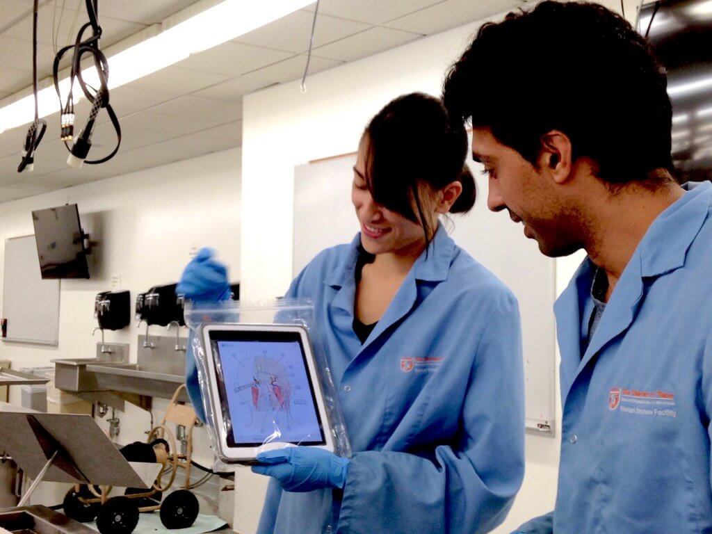
The iPads’ predecessor, standing computers, offered students and educators a limited range of use and communicability during dissections and lab lectures. Computer stations were used in the gross anatomy lab, the area in medical schools dedicated to studying organs and tissues at the macroscopic level, for the past three years, but that proved to be difficult for students. Now, each of the 42 cadaver tanks in the lab is equipped with an iPad, protected with a shock-proof case wrapped in a Zip-Lock bag to prevent unwelcome material from damaging the expensive pieces of hardware and from getting — to use the double entendre — gross.
According to Cleary, students use the iPads frequently and find them beneficial for many of the same reasons people generally like them to begin with. The tablets’ mobility and interactive interface allows each student in the standard groups of three to be involved and active participants.
“Our group really enjoys using the iPad because you can’t have three different sets of hands working on the cadaver at the same time, so one person is usually directing [with the iPad] and saying, ‘You need to be cutting this or removing that,’” first-year medical student Kendall Masada said.
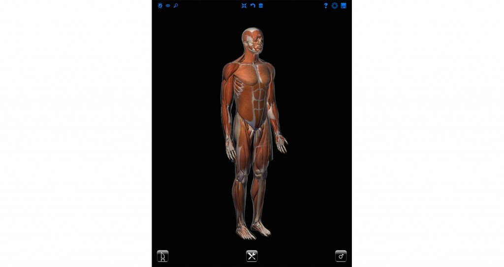
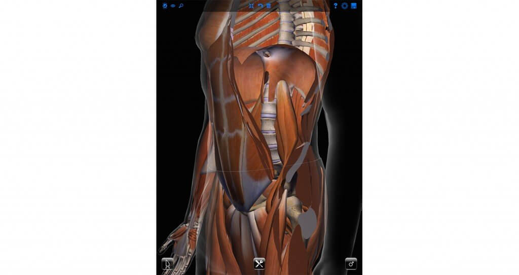
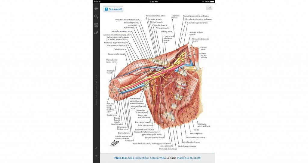
Students with the iPads also have digital access to searchable information from the internet and libraries at the tips of their fingers, a feature that is especially helpful when students are still learning and memorizing all of the different structures and intricate pieces of human anatomy. Though some students still opt for physical hard copies of books, the tablet doubles as an e-reader that students can use to access 3-D rotating models of the human body and digital versions of their textbooks, dissecting manuals and cadaveric atlases.
“Where the benefit of it lies is that you have so many resources that are pretty quick to access,” first-year medical student Yaqub Betz said. “Going through your textbook takes a long time and you’re going to get that textbook really messy. You can use the iPad to find certain structures you don’t understand and go through some of the dissection instructions if you’re not very familiar with something.”
Other students have enjoyed working with the iPads because of the device’s camera features. With the iPad’s mobility and its eight-megapixel front camera, students use the tablet to take close-up, high-definition photos that give them better visuals of their specimen that they wouldn’t be able to see with the naked eye.
“I’ll zoom in [to take a picture] and turn the iPad around to my lab partner to show them which area they’re supposed to be looking at,” Masada said.
“You can physically take and put it somewhere or turn it around,” first-year medical student Alissa Chen added. “If [someone] is dissecting, I can hold it up for her and she can simply look up to see the screen, instead of looking around for [it].”
Cleary said the iPads have been proven to be a useful and informative platform for the students’ curricula and learning experience, but the devices also add an element of fun to the lab exercises. The iPads are installed with apps that allow students to quiz each other about anatomical structures and concepts.
By applying the same game design and interactivity concepts as other tablet games, “You remove all the labels and show the picture to someone. You try to compare it to your cadaver and say, ‘It think that’s the nerve,’” fellow first-year medical student Pedram Honarpisheh said. “It’s a very noninvasive integration of the [tablets]. It doesn’t interfere at all and it just adds fun, too.”
Cleary said these capabilities are just the beginning though: Incoming first-year students can soon look forward to more mobile technology in the lab, including the ability for students to share iPad screens with larger monitors and to construct their own atlases by compositing pictures taken with the integrated cameras.